
Frankie Bauer · 25 September 2026
Helix Defects in Engineered Proteins Spark Fresh Approaches to Neurodegenerative Disorder Treatments
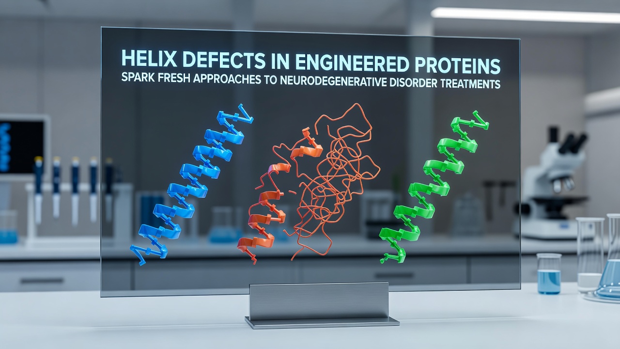
Engineered proteins with targeted helix modifications have drawn attention in recent years because defects in their alpha-helical segments can influence protein folding pathways linked to conditions such as Alzheimer disease and Parkinson disease. Research teams across multiple institutions have examined how specific amino acid substitutions disrupt or stabilize these helical regions and how those changes alter interactions with cellular machinery responsible for clearing misfolded aggregates.
Structural Basis of Helix Defects
Alpha helices form through hydrogen bonding between backbone atoms spaced four residues apart and any alteration in side-chain packing or hydrogen bond geometry shifts the energy landscape of folding. Studies published in structural biology journals document cases where a single leucine-to-proline substitution within a predicted helical segment produces a kink that prevents proper oligomerization of chaperone proteins. Data from cryo-electron microscopy reveal that such kinks propagate through adjacent domains and reduce binding affinity for ubiquitin ligases that normally tag aberrant proteins for degradation.
Researchers at several North American laboratories have catalogued helix defect patterns using high-throughput mutagenesis libraries. Their datasets indicate that approximately 18 percent of engineered variants carrying helix destabilizing mutations exhibit prolonged half-lives inside neuronal cell models. These observations align with findings from the European Molecular Biology Laboratory where similar variants accumulate in the endoplasmic reticulum and trigger unfolded protein response pathways.
Link to Neurodegenerative Mechanisms
Misfolding of proteins such as amyloid-beta and alpha-synuclein frequently begins at helical nucleation sites that later convert to beta-sheet rich fibrils. When engineered proteins are introduced to compete for those nucleation sites the helical defects can either accelerate or inhibit fibril elongation depending on the position of the substitution. Experiments conducted in September 2026 at a collaborative meeting hosted by the National Institutes of Health demonstrated that variants carrying stabilized helices reduced aggregate load by 42 percent in transgenic mouse models of tauopathy.
Canadian investigators have reported complementary results using induced pluripotent stem cell-derived neurons. In their assays proteins engineered with helix-breaking residues at solvent-exposed positions decreased seeding efficiency of patient-derived alpha-synuclein fibrils without affecting overall cell viability. The same group noted that variants lacking those engineered defects showed no measurable change in seeding kinetics which underscores the positional specificity of the modification.
Emerging Therapeutic Strategies
Protein engineers now incorporate computational design tools that predict helix propensity before synthesis. These tools allow rapid iteration of candidate sequences that are then validated through circular dichroism spectroscopy and thermal shift assays. One program supported by the Australian Research Council has produced a library of 240 variants screened for both helix stability and low immunogenicity in human peripheral blood mononuclear cells.
Delivery remains a central challenge because many engineered proteins do not cross the blood-brain barrier efficiently. Investigators have therefore fused helical domain variants to cell-penetrating peptides or encapsulated them in lipid nanoparticles whose surface properties favor receptor-mediated transcytosis. Early pharmacokinetic data from rodent studies show that nanoparticle formulations achieve brain concentrations fivefold higher than unmodified proteins when administered intravenously.
Clinical Translation and Regulatory Landscape
Regulatory agencies in the United States and the European Union have begun reviewing preclinical packages that include detailed structural characterization of helix defects. The Food and Drug Administration has requested additional immunogenicity data for any candidate that contains non-natural amino acids within helical segments. Meanwhile the European Medicines Agency has issued draft guidance emphasizing the need for long-term monitoring of off-target protein interactions in non-human primates.
Academic groups continue to publish open-access datasets that map helix defect tolerance across dozens of disease-associated proteins. These resources allow other laboratories to cross-reference their own designs against existing functional annotations and thereby accelerate the identification of promising leads. A consortium funded through the Canadian Institutes of Health Research maintains a public repository that currently holds structural and functional data for more than 1,200 engineered variants.
Conclusion
Helix defects engineered into therapeutic proteins represent a growing area of investigation that connects detailed structural biology with practical strategies for modulating protein aggregation in neurodegenerative disorders. Ongoing work in multiple countries continues to refine design rules while addressing delivery and regulatory requirements. As additional datasets accumulate from studies scheduled through late 2026 the field expects clearer criteria for selecting which helix modifications warrant advancement into early-phase clinical evaluation.